Pages

Showing posts with label skull. Show all posts

Thursday, December 16, 2010

Letter of the Day: December 16

Beuchene Skull aka "Exploded" Skull

Curatorial Records: Numbered Correspondence 1872


Mssrs Richard Kny & Co.,

17 Park Place,

New York.


Gentlemen:


Will you have the kindness to inform me at an early date of the prices at which you will furnish the following preparatioins:


Osteological Preparations, Catalogue III

p. 3. Skull disarticulated, mounted according to Beauchene, bones untied by polished metal strips, screw movement.

p. 3 Ear with two cuts, internal and median.

p. 15. Skull of monkey, mounted after Beauchene.


Biological Preparations, Catalogue No. V, pp. 20 and 21.


I.                    Dissected Preparations.

Mus decumanus

Columba sp.

Lacerta agilis.

Rana fortis.

Tinca vulgaris.

Bombyx mori.

Astacus fluviatilis.

Helix pomatis.


II.                  Injected preparations.

Mus decumanus.

Columba sp.

Lacerta agilis.

Rana fortis.

Tinca vulgaris.

Astacus fluviatilis.

Helix pomatis.

Hiruda medicinalis.


III.                Nerve Preparations.

Mus decumanus.

Columba sp.

Lacerta agilis.

Rana escuelenta.

Melolontha vugaris.

Hydrophilus piceus.

Astacus fluviatilis.


Very respectfully,


D.L. Huntington

Deputy Surgeon General, U.S. Army,

In charge of Mus. and Lib. Div.


Additional

Hiruda medicinalis, nerve prep.

Helix pomatis, “ “


Monday, September 6, 2010

Letter of the Day: September 6

Dear Doctor:

I have been called upon to give testimony in a criminal case - in which there is a bullet wound of the head with extensive fracture of the skull – and no external marks of violence.

Would I be asking or troubling you too much in requesting you to send me such photographs as will have a bearing upon the case, such as will illustrate the average amount of fracture of the skull from bullets, + especially pistol shots.

Also such as will illustrate well authenticated cases of fracture from “Contre Coup.”

With great respect
I have the honor to be-
Very Sincerely,
Your Obdt Servant
A. Van Deveer

^^^^

Respectfully submitted to the Surgeon General, U.S.A. for instructions. A certain number of the illustrations contained in the Army Medical Museum on the subjects referred to have been photographed and prints have been furnished to two medical men of Albany, engaged in a medico-legal inquiry – possibly the same to which Dr. Van Derveer refers.

George A. Otis
Ass’t Surg. USA

SGO
Sept. 13. 69.

^^^^

Let him have them if in hand --

Sunday, July 18, 2010

Letter of the Day: July 18 - French skull

Curatorial Records: Numbered Correspondence 44

July 18, 1894

Mr. Gustav Goldman,
Maryland General Hospital,
Linden Ave., North of Madison St.,
Baltimore, Md.

Dear Sir:

In reply to your favor, just received, I beg to inform you that a French skull, disarticulated, will be forwarded to your address this afternoon.

Hoping that it will prove satisfactory, I remain,
Yours very sincerely,
Walter Reed
Major and Surgeon, U.S. Army,
Curator Army Medical Museum.

The skull has been sent as directed, to 841 Hollins St., Baltimore

Tuesday, June 17, 2008

Mememto Mori, or, The Head of Janus


I made a rewarding visit to the Musée d'Histoire de la Médecine in Paris last month, but thoroughly grossing out my husband with its extensive exhibits of surgical tools, including kidney stone extractors. Not something that a man who has had a kidney stone especially wants to look at. I've not had kidney stones, and I found the tools and illustrations painful to look at, and I've seen some pretty gross things in the course of my job. Do you want to see them? Next time.

I really liked this ivory carving from the 17th century, called the Head of Janus. I don't know if the Catholic school I went to didn't teach mythology as a matter of theology or what, but I never learned about the myths. So wikipedia to the rescue: "Janus was usually depicted with two heads (not faces) looking in opposite directions, and was frequently used to symbolize change and transitions such as the progression of past to future, of one condition to another, of one vision to another, the growing up of young people, and of one universe to another. He was also known as the figure representing time because he could see into the past with one face and into the future with the other."

There's nothing that says transition from one condition to another like a face on one side and a skull on the other.

Wednesday, May 7, 2008

Reeve233


Reeve233
Originally uploaded by otisarchives3
This has been a pretty big hit over the last couple of days, so we figure some blog must have linked to it. The wages of sin, y'all.

Tuesday, April 22, 2008

St. Elizabeth's Calvarium - Dr. I.W. Blackburn

A good day at the museum is rediscovering the history of a specimen or artifact that has lost its association with the record that tells us who, where and why it has come to the museum. Sometimes it takes archival research to do this and sometimes it's purely serendipitous.

This weekend I discovered a copy of "Intracranial Tumors Among the Insane (1902) by Dr. I. W. Blackburn in a used bookstore in Gaithersburg, MD. Dr. Blackburn was the former pathologist for St. Elizabeth's Hospital in Washington, D.C. He performed hundreds maybe thousands of autopsies on patients who died at the hospital. While browsing through the book I noticed a photo of very unique calvarium (top of the skull). The specimen had two rare conditions; scaphocephaly and hyperostosis frontalis interna. The bone looked strangely familiar.

In the Anatomical Division of NMHM we have such a specimen. It was listed as coming to the museum from an early exchange with the Smithsonian National Museum and not attributed to St. Elizabeth's at all. I bought the book for $15 and lo and behold when I brought it back to the museum our specimen was the same one in the book. It was attributed to a 65 year old black female patient at St. Es. The existing record was based on a bygone curatorial staff member using the wrong numbering system to describe the specimen. There have been several systems in place at the museum at various times which causes a lot of confusion for us today.

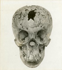
Here is a recent photo of the specimen. In addition to the pathological conditions there are also consistencies among the size and shape of the exposed frontal sinus, the etchings of the meningeal vessels, the contours of the thickened frontal bone and the two small bony exostoses in the center just left of the midline. The front of the skull is oriented to the right.

The specimen's history is now restored. Additionally, four other calvaria in the collection with no known history have similarly composed autopsy numbers written on the bones. All are now believed to be from St. Elizabeth's with further research pending. These specimens have very early accession numbers which means that they arrived at the museum around 1917-1918 when the Army Medical Museum was busy attending to the medical needs of World War I. It is not clear when the original error was made, but it likely extends back several decades. The specimens themselves are from the late 19th century autopsies.

In the photo below you can see the scaphocephalic calvarium (left) next to a normal one (right). Notice that the normal one on the right has a jagged line called the sagittal suture (front to back) which the one on the left lacks. Sutures are where the bones of the cranium grow and expand. In scaphocephaly the sagittal suture fuses prematurely and the coronal suture continues to grow which gives the unique elongated shape you see here. The one on the left is darker due to over 100 years of dust and dirt adhering to oils that remained in the bone. Since bone is porous, it can absorb materials from the environment which effect its color. The one on the right was cleaned using chemicals that removed much of the oils and was stored in a relatively cleaner environment.